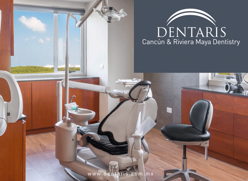
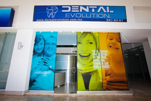
Dental Evolution is a primary Dental Clinic in Cancún, Dentaris is one of the oldest dental care centers in Cancun, having placed over 7000 implants from 1993 until present, Coral Dental Center is the first specialized dental care clinic in the Mayan Riviera.
Their teams of highly qualified professionals have the latest equipment and technology to provide an efficient, quality service in several dental specialties. The main principal is to provide painless treatment in an atmosphere of comfort and tranquility.
Their locations allow them to care for not only patients living in the area (Playa del Carmen, Cancun, Cozumel, Merida) but also to patients from all over the world who decide to take a dental vacation in paradise.
Instruments that do not resist high temperatures, such as plastics, are disinfected in a chemical solution that kills those same infectious micro-organisms.
Many implements are disposable and are only used for one patient. This includes all needles and aspirators (the plastic straw placed in the mouth to suck up saliva).
Gloves and masks are used to prevent disease transmission. Gloves are never used for more than one patient.
All surfaces are disinfected with a chemical solution specially designed to kill bacteria, spores and viruses. Protection barriers (plastics, rubber drapes, etc) are used whenever possible.
We constantly monitor our procedures in order to meet the OSHA infections control and the recommendations of American Dental Association.
The following procedures are performed: | |
Dental Examination | |
Dental Implantology | |
Orthodontic Treatment | |
Dental Whitening | |
Pediatric Dentistry | |
Night Guard | |
Basic Cleaning | |
Restorative Dentistry | |
Cosmetic Dentistry | |
Temporary crowns |
Your Doctor will recommend a recovery time in the hospital or give you further instructions on how to go through the next days in your hotel and when to come back for further checks.
Your Medical Concierge will get all needed pharmacy shopping done. He will help you through the recovery process.
The only thing you have to do is to get better.
Regular dental examinations (ideally every six months) are very important and help you to maintain a healthy mouth. The process allows the dentist to check for early signs of dental problems, and therefore take measures to prevent the problems from becoming serious. This could save a lot of pain, time and money!
What is involved in a Dental Examination?
FAQS:
Why should I apply for a dental examination?
Regular dental exams are an important part of preventive health care. During a dental exam, the dentist or hygienist will clean your teeth and identify gum inflammation or bone loss. The dentist or hygienist will evaluate your risk of developing tooth decay and other oral health problems.
What Should I Expect from a Routine Dental Cleaning?
A dental cleaning is a fairly common procedure that is rarely painful. Those who fear dentists, or who have marked tooth sensitivity may feel more discomfort than the average patient. Similarly, those who have not had their teeth cleaned for a long time, expecting to experience more cleaning and some additional pulling or scraping to remove plaque from your teeth.
How Often Should You Go to the Dentist?
Dental and health organizations decided there was a need to set standards for preventive dentistry. Even if you take excellent care of your teeth and gums at home, you still need to see your dentist regularly. Regular visits allow your dentist to find early signs of disease.
Do bad teeth lead to bad general health?
There is significant research being undertaken which suggests that there is a link between periodontal disease (gum disease) and heart disease. It is proposed that local periodontal inflammation can result in a wider immune response in the body which leads to a response in the lining of arteries facilitating atherosclerosis and heart disease.
What Does a Typical Dental Examination Include?
A number of factors should be evaluated in a typical dental examination, including soft tissue condition, periodontal status, existing cavities and fillings, complete set of x-rays and general appearance of smile.
Is a root device usually made of titanium used in dentistry to support restorations that resemble a tooth or group of teeth to replace missing tooth.
First, you will need to discuss your options with your dentist. Together, it will be decided if you are a good candidate for dental implants. The dentist will take a complete dental history, x-rays, and complete a thorough oral examination.
If you are a candidate for implant surgery, the procedure is as follow:
Surgical placement of the implant into the bone (usually in the dentist office).
There is a healing period of approximately four months. (Osseointegration: the implant fuse to the bone).
Next, there is a minor surgical exposure of the top of the implant.
The last phase is the restorative phase. The dentist will take impressions then make a prosthesis that will attach to the implants. This will require several visits.
Some factors will contribute to long-term success of dental implants (bone, habits, surgeon, rehabilitation, cleaning, dental examination etc). And Dental implants have given countless patients many amazing benefits.
FAQS:
WHAT IS AN IMPLANT?
Implant is a screw made of pure titanium which has been subjected to a special surface treatment to ensure the bone osseointegration. Is a biocompatible medical prosthesis, suitable to be implanted in the human body, an implant is a man-made replacement for natural teeth which allows the person to return to fixed teeth. It is not a transplant, which is taken from another person. There are several categories of dental implants, which will be selected by the doctor depending on your specific needs and general dental condition.
Are there any age limitations for dental implants?
No. Any person at any age can have dental implants as long as there is enough bone available in which to place the implants.
How often will I need to have my dental implants checked?
The success of your implants will depend greatly on how well you maintain them. They will need to be professionally cleaned by a hygienist and examined by your implant dentist every three to four months.
Is dental implant surgery painful?
No. An effective local anesthetic is used during the surgery so that you do not have any discomfort during the placement of the implants. The mild discomfort you might experience after surgery can be controlled with medications.
How long can implants last?
Different long term research studies have shown that implants which have been placed in good bone can last for a patient's lifetime. However, some implants can fail if the quality of the bone wasn't good, if there was too much bite stress for too few implants or implants which weren't long enough for the demands of a given case.
Orthodontic treatment is used to correct a bad bite or malocclusion, which involves teeth that are crowded or crooked. In some cases, the upper and lower jaws may not meet properly and although the teeth may appear straight, the individual may have an uneven bite.
Protruding, crowded or irregularly spaced teeth and jaw problems may be inherited. Thumb-sucking, losing teeth prematurely and accidents also can lead to these conditions.
Correcting the problem can create a nice-looking smile, but more important, orthodontic treatment results in a healthier mouth. That’s because crooked and crowded teeth make cleaning the mouth difficult, which can lead to tooth decay, gum disease and possibly tooth loss. An improper bite can interfere with chewing and speaking, can cause abnormal wear to tooth enamel, and can lead to problems with the jaws.
Braces (also called orthodontic appliances) can be as inconspicuous or as noticeable as you like. Brackets the part of the braces that attach to each tooth are smaller and can sometimes be attached to the back of the tooth, making the brackets less noticeable.
Brackets may be made of metal, ceramic, plastic or a combination of these materials. Some brackets are clear or tooth-colored. There are brackets shaped like hearts and footballs, and elastics (orthodontic rubber bands) in school colors or holiday.
Malocclusions often become noticeable between the ages of 6 and 12, as the child’s permanent (adult) teeth erupt. Orthodontic treatment often begins between ages 8 and 14. Treatment that begins while a child is growing helps produce optimal results. As a result, children should have an orthodontic evaluation no later than age 7. By then, they have a mix of primary (baby) teeth and their permanent (adult) teeth. Your child’s dentist can spot problems with emerging teeth and jaw growth early on, while the primary teeth are present. That’s why regular dental examinations are important.
Children aren’t the only ones who can benefit from orthodontics. If you’re an adult, it’s not too late to correct problems such as crooked or crowded teeth, overbites, under bites, incorrect jaw position, or jaw-joint disorders. The biological process involved in moving teeth is the same at any age. Usually, adult treatment takes a little longer than a child's treatment. Because an adult's facial bones are no longer growing, certain corrections may not be accomplished with braces alone. No matter your age, it's never too late to improve your dental health and beautify your smile.
Orthodontics is a specialty area of dentistry that is officially known as Orthodontics and Dentofacial Orthopedics. The purpose of orthodontics is to treat malocclusion through braces, corrective procedures and other "appliances" to straighten teeth and correct jaw alignment. An orthodontist is a dentist who specializes in the diagnosis, prevention, and treatment of dental and facial irregularities.
Although treatment plans are customized for each patient, most wear their braces from one to three years; depending on what conditions need correcting. This is followed by a period of wearing a "retainer" that holds teeth in their new positions. Although a little discomfort is expected during treatment, today’s braces are more comfortable than ever before. Newer materials apply a constant, gentle force to move teeth and usually require fewer adjustments. Patients with braces should maintain a balanced diet and limit between-meal snacks.
FAQS:
If my teeth have been crooked for years, why do I need orthodontic treatment now?
There’s no time like the present, and healthy teeth can be moved at any age. Orthodontic treatment can create or restore good function, and teeth that work better usually look better, too. A healthy, beautiful smile can improve self-esteem, no matter your age.
Do I need to change my oral hygiene routine during orthodontic treatment?
Yes, keeping your teeth and braces (or other appliances) clean requires a little more effort on your part. Your orthodontist will explain how to brush and floss, how often to brush and floss, and give you any special instructions based on the kind of orthodontic treatment you are having.
In general, patients with braces must be careful to avoid hard, sticky, chewy and crunchy foods. They should also avoid chewing on hard objects like pens, pencils and fingernails.
What are my options if I don't want braces that show?
Should your case warrant it, you might want to ask your orthodontist about lingual braces, which are attached behind the teeth. Ceramic braces may be another option to lessen the visibility of braces; they blend in with the teeth for a more natural effect.
How long will orthodontic treatment take?
The average case of orthodontic treatment takes about 18-24 months, but every case is different. More severe cases could take longer. But the better you look after your brace and regularly attend appointments, the more chance you will have of a speedy treatment.
Does wearing a brace hurt?
It’s probable that when you first have your brace fitted and then come back for adjustments that you may feel some discomfort. However, this should only last a few days and you can take a mild painkiller if necessary.
Is one of the most popular cosmetic dentistry treatments to improve and invigorate the smile and is used to correct discoloration of the teeth by removing the brown and yellow staining (from the effects of certain beverages and foods: red wine, medication, coffee, smoke, and tea are the most common culprits).
For many patients, this procedure is just the first step in remaking a smile with the skill and artistry of cosmetic dentistry. Following the teeth whitening treatment, newly placed all- porcelain crowns, porcelain veneers and / or dental bonding material are matched perfectly to the new lighter color creating an amazing smile.
The procedure can be performed entirely in the cosmetic dentist's office or it can be performed by the patient in their home, using a materials provided by the dentist.
LASER WHITENING PROCEDURE
We prepare the mouth for treatment.
Really important to make a dental cleaning.
Checking the color before treatment.
Place a soft holder to gently hold back the lips during treatment.
Apply a special gel to protect the gum tissue.
Apply a special bleaching gel on the teeth that will react with the special LASER.
Reapply bleaching gel to the teeth every 20 minutes for about an hour—until the session is complete.
Then remove protective gel from the gums and removes bleaching gel from the teeth.
We present before-and-after color to the patient to view the results.
FAQS:
What is a teeth whitening?
Is the process of bleaching your teeth in order to make them appear whiter, teeth whitening is a purely cosmetic procedure, done to enhance ones smile. There are many over-the-counter teeth whitening products which have become very popular lately.
Is teeth whitening safe?
Teeth whitening are a great way to improve one’s appearance, particularly the smile. The best candidate is the person who regularly visits the dentist and does not have any signs that additional work is necessary.
How long do the teeth whitening effects last?
People who expose their teeth to foods and beverages that cause staining may see the whiteness start to fade in as little as one month. Those who avoid foods and beverages that stain may be able to wait one year or longer before whitening treatment or touch-up is needed.
Is teeth whitening painful?
Teeth whitening are not supposed to be painful, but with that said keep in mind that it might cause tooth and / or gum sensitivity. If this happens it's a good idea to discontinue the treatment for a while and talk to dentist about the issue.
What is laser tooth whitening?
The real-life procedure pales in comparison to the process envisioned by the overactive imagination. Repeated applications and expensive equipment that may or may not work the process can be done in a single session. It also, unlike many other dental procedures does not result in a great deal of pain.
Since children begin to get their baby teeth in the first several months of life even infants should undergo an oral exam. This exam includes a risk assessment for caries (an infectious disease) in both baby and mother.
The pediatric dentist strives to make his office a fun place for risk to be taught about proper oral hygiene. They perform preventive care such as teeth cleaning and fluoride treatments as well as providing dietary recommendations, treat tooth decay, make pulpotomies , temporary crowns , healings , extractions ,can also offer young children an early evaluation and treatment for the straighten of teeth (orthodontics).
Another job of the pediatric dentist is to diagnose oral abnormalities that may be connected to other problems including asthma, diabetes, congenital heart defects, etc.
There are several gum diseases and conditions that pediatric dentists treat and manage. They include pediatric periodontal disease and ulcers.
Pediatric dentists also care for injured teeth that may have fractured, been displaced or knocked out.
FAQS:
When should I take my child to the dentist for the first check-up?
In order to prevent dental problems, your child should see a pediatric dentist when the first tooth appears or no later than his/her first birthday.
How often does my child need to see the pediatric dentist?
A check-up every six months is recommended in order prevent cavities and other dental problems. However, your pediatric dentist can tell you when and how often your child should visit based on their personal oral health.
What should I use to clean my baby's teeth?
A toothbrush will remove plaque bacteria that can lead to decay. Any soft-bristled toothbrush with a small head, preferably one designed specifically for infants, should be used at least once a day at bedtime.
Why Are The Primary Teeth So Important?
It is very important to maintain the health of the primary teeth. Neglected cavities can and frequently do lead to problems which affect developing permanent teeth.
What’s the best toothpaste for my Child?
Many toothpastes, and / or tooth polishes, however, can damage young smiles. They contain harsh abrasives which can wear away young tooth enamel. When looking for toothpaste for your child make sure to pick one that is recommended by the American Dental Association.
Bruxism is the clenching together of both the upper and lower teeth. Clenching can cause pressure on the muscles, tissues, and other surrounding structures of the jaw, and can cause jaw joint disorders, headaches, neck pain, jaw pain, ear pain, tense muscles, and excessive wear on the teeth.
A large number of people who clench also tend to grind their teeth. Grinding is when you slide your teeth over each other in a back and forth, sideways movement. Often the grinding is a subconscious behavior that is not realized by the bruxer.
Stress reduction and anxiety management are methods used to treat bruxism and reduce the symptoms, but a dental night guard is the most commonly used treatment method.
A night guard is a horseshoe-shaped, retainer-like, plastic appliance with shallow borders that can be worn on either the top or the bottom arch. It is designed to protect the teeth from the pressure of clenching and grinding by providing a protective cushion between the upper and lower teeth.
A night guard can be a simple and affordable solution for protecting your teeth and jaws from the damaging effects of bruxism. The procedure is really simple; we take an impression, send it to the laboratory and are ready the next day.
Dental cleanings involve removing plaque (soft, sticky, bacteria infested film) and tartar (calculus) deposits that have built up on the teeth over time.
The purpose of the cleaning and polishing is basically to leave the surfaces of the teeth clean and smooth so that bacteria are unable to stick to them and you have a better chance of keeping the teeth clean during your regular home care.
We commonly used an ultrasonic instrument which uses tickling vibrations to knock larger pieces of tartar loose. It also sprays a cooling mist of water while it works to wash away debris and keep the area at a proper temperature.
Then using a jet formed by a mixture of air, powder and water, the AIR-FLOW removes dental plaque, soft deposits and surface stains from pits, grooves, interproximal spaces and smooth surfaces of the teeth.
Polishing is done using a slow speed hand piece with a soft rubber cup that spins on the end.
Most people find that cleanings are painless, and find the sensations described above – tickling vibrations, the cooling mist of water, and the feeling of pressure during "scraping" – do not cause discomfort.
Painful cleaning experiences can be caused by exposed dentine (not dangerous, but can make cleanings unpleasant), or sore gum tissues.
FAQS:
What is basic dental care?
Basic dental care involves brushing and flossing your teeth regularly, seeing your dentist and / or dental hygienist for regular checkups and cleanings, and eating a mouth-healthy diet, which means foods high in whole grains, vegetables and fruits, and dairy products.
Why is basic dental care important?
Why should I apply for a basic cleaning?
Taking care of your teeth with basic cleanings and active dental care can help to prevent many painful and costly problems in the future. It's all about prevention. Brush your teeth often, floss your teeth well, and get your teeth cleaned professionally at the dentist's office at the appropriate intervals.
Why is it vital to visit the dentist for periodic cleanings?
First, that makes it easier for your dentist to ensure the health of your teeth. Cavities can be detected and repaired promptly. Second, and just as importantly, regular cleanings remove plaque and tartar that not only detract from the luster of your teeth but also eat away at your dental health, promoting tooth decay, bad breath and gum disease.
What are the different types of dental cleanings?
There are two types of dental cleanings: standard teeth cleaning and deep teeth cleaning. Dentists recommend standard teeth cleaning twice a year, though it is sometimes performed more often as part of an on-going dental treatment. Deep teeth cleaning are more complex and performed only when it is necessary.
Restorative Dentistry
Is a dental restorative material used to restore the function, integrity and morphology of missing tooth structure?
In dentistry, crown and bridge (Restorative Dentistry) refers to the restoration of natural teeth that have been damaged, decayed or lost. Once your dentist has examined your teeth and has evaluated your dental and medical history, he/she will be ready to provide a diagnosis, and treatment options. A crown may be constructed to restore an individual damaged tooth back to its original form and function, while a bridge may be utilized to replace one or more teeth. These restorations are cemented onto the teeth and are referred to as "fixed" dentistry as opposed to a restoration of missing teeth with a removable appliance or partial denture.
RESTORATION CLASSIFICATIONS: depending on their size and location we use different kind of materials and techniques.
FAQS:
What is Restorative Dentistry?
Restorative dentistry replaces old mercury fillings, old ugly crowns and composites that have become discolored. Restorative dentistry also repairs cracked teeth and / or missing teeth.
Who would benefit from Restorative Dentistry?
Restorative Dentistry is ideal for an individual wanting to renew restorations in a couple easy visits to ensure overall oral health and a beautiful smile that feels good. He or she may be tired of "piece-meal" dentistry that never seems to quite solve a problem with a tooth.
How long does Restorative Dentistry take?
Restorative Dentistry is usually accomplished in two visits each ranging from 2 to 4 hours depending on the procedure.
Will my teeth look natural after a restorative dentistry?
Yes, sure. Harmony will be achieved between your existing teeth and the restored ones. It is not only achieved on the teeth level, but also between your gums, lips, and your face in general.
Will I have to do extra effort for cleaning my restored teeth?
It will be the same effort that you do to clean your natural teeth. You will need to brush and floss them regularly.
REAL SMILE WITH VEENERS
VENEERS: IMPORTANT TOOL FOR THE COSMETIC DENTIST TO CREATE A "HOLLYWOOD" TYPE OF MAKEOVER is a thin layer of restorative material placed over a tooth surface. We may use to restore a single tooth that may have been fractured or discolored , close spaces, lengthen teeth that have been shortened by wear, provide a uniform color ( teeth that do not respond well to whitening procedures) , shape, and symmetry, and make them appear straight.... in conclusion "we can created exquisite smile makeovers with veneers "
We can use two can of material for veneers ; "composite " and "porcelain"
Composite veneers are more prone to staining and do not last as long as porcelain veneers. Porcelain veneers also offer a more natural looking, translucent appearance, the procedure of having dental veneers fitted is:
Initial consultation and examination
The first stage is to numb the teeth and gums with a local anesthetic so that you do not feel anything during the procedure.
Using a special tool called a burr (a dental drill or file); a tiny part of the front surface of your teeth will be shaved off.
The amount removed should be equivalent to the thickness of the veneer that will sit over the top of the tooth (.5mm), next an impression or mould (copy) of your teeth is taken you will use a temporary veneer for some days.
After you come back just for fitting the permanent veneer (checking color, form etc...).
FAQS:
What is cosmetic dentistry?
Cosmetic dentistry is dental treatment that improves the beauty and health of one's smile. Nearly every dental procedure has 2 elements: Aesthetics and function. Quality cosmetic dentistry takes both of these elements into consideration; so that one may rest assured they will not only look great, but enjoy hassle free long-term results.
What are some examples of cosmetic dentistry procedures?
Cosmetic dentistry covers a multitude of procedures from smile teeth whitening to repairing, straightening, replacing or enhancing the appearance of one’s teeth and smile. Most common is the use of Cosmetic Veneers or Laminates. These thin, but durable restorations are much more conservative to the natural tooth than traditional crowns, and have become a very popular way to enhance a smile.
Why would someone need or be interested in cosmetic dentistry?
Cosmetic dentistry provides more than just a beautiful smile and a healthy mouth. According to the American Academy of Cosmetic Dentistry (AACD), cosmetic dentistry has also been shown to improve a person's overall health and emotional well-being.
How can Cosmetic Dentistry change my life?
Can a new smile make you more attractive, give you improved confidence, a healthier attitude, more energy, more alive? What do you think? A new smile can give you a whole new level of confidence, both socially; personally and even improve your career.
What is a cosmetic veneer?
Modern metal-free dentistry has evolved to a point where we can be much more conservative with tooth preparation. A veneer is essentially a conservative crown. Rather than encompassing the entire tooth, a cosmetic veneer, essentially covers the visible part of a tooth preserving a large amount of the natural tooth structure. Veneers can straighten, lengthen and whiten teeth to provide the desired smile and bite; each veneer tooth must be shaped.
Crowns are made of acrylic and are used as a temporary restoration until a permanent crown is constructed by the dental laboratory. However they are not as strong as the permanent and the patient has to be very carefully. These crowns also can be individual or a complete bridge, sometimes the patient wants to use it for more time and although this are less expensive they are more prone to fractures and wear down over time.
There are 2 ways for making temporary crowns it depends of the time and the patient requirements.
The first option the temporary crown can be made in the dentist’s office and the second option your dentist send it to the laboratory so it could be more esthetic and has a better seal.
FAQS:
What is a temporary crown?
A dental crown is a tooth-shaped "cap" that is placed over a tooth covering the tooth to restore its shape and size, strength, and / or to improve its appearance.
The crowns, when cemented into place, fully encase the entire visible portion of a tooth that lies at and above the gum line.
How Should I Care for My Temporary Dental Crown?
Because temporary dental crowns are just that, a temporary fix until a permanent crown is ready, most dentists suggest that a few precautions be taken with your temporary crown. Treatment includes:
Avoid sticky, chewy foods (for example, chewing gum, caramel), which have the potential of grabbing and pulling off the crown.
Minimize use of the side of your mouth with the temporary crown. Shift the bulk of your chewing to the other side of your mouth.
Avoid chewing hard foods (such as raw vegetables), which could dislodge or break the crown.
Slide flossing material out-rather than lifting out-when cleaning your teeth. Lifting the floss out, as you normally would, might pull off the temporary crown.
Why Is a Dental Crown Needed?
A dental crown may be needed in the following situations:
To protect a weak tooth (for instance, from decay) from breaking or to hold together parts of a cracked tooth.
To restore an already broken tooth or a tooth that has been severely worn down.
To cover and support a tooth with a large filling when there isn't a lot of tooth left.
To hold a dental bridge in place.
To cover misshapen or severely discolored teeth.
To cover a dental implant
What if my temporary crown comes off?
Most important thing is not to panic if this happens; this is not an emergency situation, but do not be surprised if the exposed tooth is a bit sensitive. The temporary crown does need to be re-cemented soon, but in the meantime follow these suggestions.
Take some petroleum jelly, toothpaste, or some DENTAL temporary cement you buy at your local pharmacy and coat the inside of your temporary crown. Place it back on your prepared tooth. It will only fit one way, so don’t worry about getting it on incorrectly.
It is very important to wear your temporary crown at least several hours each day in order to maintain the spacing. If you do not wear it some time each day, the permanent crown may not fit, and the procedure will have to be redone.
What happens if I swallowed a temporary crown?
If you know for a fact you've swallowed the item in question then you have a choice to make. Do nothing and the item will pass harmlessly through your innards and end up in the city's water-treatment facility. On the other hand, it is possible to recover the item. It usually takes just 24-48 hours. Yes, if you are willing to do what's necessary the item will reappear after its Fantastic Journey and can be cleaned sterilized and (in the case of a crown) re-cemented!
Microlaminectomy is performed for patients with symptomatic, painful lumbar spinal stenosis. It is performed to remove the large, arthritic osteophytes (bone spurs) that are compressing the spinal nerves. This is microscopic or endoscopic surgical approach performed using a small incision, with minimal dissection, to accomplish a nerve root lumbar decompression. This minimally invasive approach allows for a more rapid recovery.
FAQS:
What is a Microlaminectomy?
Microlaminectomy is a minimally invasive surgical procedure used to treat patients suffering from bone spurs compressing the spinal nerves, lumbar spinal stenosis, or herniated lumbar discs, which are sometimes referred to as slipped discs or ruptured discs. Traditional surgery uses large incisions that cause trauma to the muscle and nerve tissue, often resulting in increased pain and a longer hospital stay. In contrast, microlaminectomy is a microscopic surgical approach that uses two-to-three centimeter-long incisions that do not damage muscle and other soft tissues.
What happened after surgery?
Most patients are usually able to go home 1-2 days after surgery. Before patients go home, physical therapists and occupational therapists work with patients and instruct them on proper techniques of getting in and out of bed and walking independently. Patients are instructed to avoid bending at the waist, lifting (more than five pounds), and twisting in the early postoperative period (first 2-4 weeks) to avoid a strain injury. Patients can gradually begin to bend, twist, and lift after 1-2 weeks as the pain subsides and the back muscles get stronger.
What is the chance of being cured?
The results of microlaminectomy surgery in the treatment of symptomatic spinal stenosis are generally excellent. Numerous research studies in medical journals demonstrate greater than 86-95% good or excellent results from microlaminectomy surgery, and often show an improved recovery time compared with patients undergoing conventional, open laminectomy surgery. Most patients are noted to have a rapid improvement of their pain and return to normal function.
Using innovative technology, a minimally invasive surgery (MIS) spinal fusion (mending the spine bones together) can now be accomplished using two small e incisions with minimal tissue dissection resulting in a faster recovery and less pain than traditional open spinal fusion surgery. Posterior Lumbar Fusion (PLF) is the general term used to describe the technique of surgically mending two (or more) lumbar spine bones together. Minimally invasive PLF is performed instrumentation (use of metal screws/rods) to impart immediate stability while the bone mends together. The MIS PLF technique is often favored when a laminectomy is not required. It is performed for a variety of spinal conditions, such as spondylolisthesis and spinal instability, among others.
FAQS:
When should I consider surgery?
Surgery should always be the last resort when it comes to treating spinal conditions in the neck and back. However, if various non-operative treatments have been attempted without improvement or worsening over a 6-12 month period, then surgical treatment seems reasonable for certain specific conditions such as spinal stenosis, sciatica, spondylolisthesis or degenerative scoliosis. The decision for surgery should be individualized to the patient and the patient’s symptoms, along with their level of function.
How long is the recovery?
Most patients are able to get up out of bed and start walking shortly after surgery, usually on the same or following day. For the first 6 weeks, the activity level is limited to walking and normal daily activities.
Most patients are encouraged to avoid heavy lifting, frequent bending, twisting or turning or climbing during the first 6 week period. After 6 weeks, patients begin a physical therapy and exercise program to achieve rapid recovery and strength. By 3 months a gradual increase in normal activities as well as the institution of low impact sporting activities can be started. At 6 weeks, all activities are begun, including sports.
Which type of surgery has a greater success rate?
At present, the long-term results of minimally invasive surgery are not well studied. These assessments are ongoing. The short term success of minimally invasive spine surgery is well established. It is clear that minimally invasive surgery allows more rapid recovery and return to work/sports. There is less post-operative pain and shorter hospital stay
Microlumbar Discectomy (MLD), is performed for patients with a painful lumbar herniated disc. Microdiscectomy is a very common for spine surgeons. The operation consists of removing the protruding portion of the disc that is compressing the nerve root. Today, spine surgeons use a microscopic or endoscopic surgical approach with a small, minimally-invasive incision, allowing for a more rapid recovery.
FAQS:
What is a microdiscectomy?
Microdiscectomy, also called Microlumbar Discectomy (MLD), is performed for patients with a painful lumbar herniated disc. Microdiscectomy is a very common, if not the most common, surgery performed by spine surgeons. The operation consists of removing a portion of the intervertebral disc, the herniated or protruding portion that is compressing the traversing spinal nerve root.
What is the chance of a successful Lumbar Microdiscectomy
A lumbar microdiscectomy is 85% - 95% successful in relieving pain in the lower back and leg. Pain relief after the surgery is generally rather rapid however in some acute instances it can take from six to eight weeks for the affected nerve to settle down. If the nerve has been pinched for quite some time the success rate is rarely 100% and there can be some residual weakness, mild tingling or pain which should be tolerable however.
What are the limitations of the Lumbar Microdiscectomy?
The major limitation of the surgery is a slight weakening of the affected ruptured disc post surgery. When a disc ruptures (herniates) within your pack a hole is created within the other ring. In order to remove the loose material and conduct the surgery the surgeon will enlarge this hole, however there is no way to repair the hole once it has been enlarged so while the surgery will be effective in removing the pain to your lower back and leg - you will be left with a permanently weakened outer ring around one of your discs. Care will need to be taken to prevent any undue stress to the disc for fear of re-herniation.
Now a day there are advance treatments by Neuroendoscopy for hydrocephalus. These procedures are done thru a 2cm incision in the skin and a 1cm diameter hole in the skull.
Disorders treatable using Neuroendoscopy:
Hydrocephalus Third ventriculostomy is indicated for non-communicating hydrocephalus where there is and obstruction on the aqueduct, and the intention is to bypass this obstruction. This procedure connects the ventricular system at the 3rd ventricle to the basal cisterns and the fluid can reach the arachnoidal granulations and be reabsorbed.
FAQS:
What does hydrocephalus means?
Hydrocephalus, also known as "water in the brain," is a medical condition in which there is an abnormal accumulation of cerebrospinal fluid (CSF) in the ventricles, or cavities, of the brain. This may cause increased intracranial pressure inside the skull and progressive enlargement of the head, convulsion, tunnel vision, and mental disability. Hydrocephalus can also cause death.
What is a Neuroendoscopy for Hydrocephalus?
Neuroendoscopy is a promising minimally invasive technique in the management of non-communicating hydrocephalus and hydrocephalus associated with brain tumors and intraventricular cystic lesions. However neuroendoscopic procedures are not without risks.
What is the treatment of this condition?
Hydrocephalus treatment is surgical, generally creating various types of cerebral shunts. It involves the placement of a ventricular catheter (a tube made of silastic), into the cerebral ventricles to bypass the flow obstruction/malfunctioning arachnoidal granulations and drain the excess fluid into other body cavities, from where it can be reabsorbed. Most shunts drain the fluid into the peritoneal cavity (ventriculo-peritoneal shunt), but alternative sites include the right atrium (ventriculo-atrial shunt), pleural cavity (ventriculo-pleural shunt), and gallbladder. A shunt system can also be placed in the lumbar space of the spine and have the CSF redirected to the peritoneal cavity (Lumbar-peritoneal shunt). An alternative treatment for obstructive hydrocephalus in selected patients is the endoscopic third ventriculostomy (ETV), whereby a surgically created opening in the floor of the third ventricle allows the CSF to flow directly to the basal cisterns, thereby shortcutting any obstruction, as in aqueduct stenosis. This may or may not be appropriate based on individual anatomy.
With the help of Stereotactic Neurosurgery Parkinson disease patients can be effectively treated. There are two types of stereotactic procedures available to treat these patients worldwide.
Deep brain stimulation (DBS)
Chronic deep brain stimulation is a rapidly emerging therapy for advanced Parkinson disease. Deep brain stimulation surgery technique involves implanting electrodes inside the deep nuclei of brain called as sub thalamus. These electrodes are then connected to IPG (Pacemaker) implanted underneath the skin below the clavicle through the connecting leads. This stimulates the deep brain nuclei, which results to regression of tremor and stiffness. With the progress of deep brain stimulation disease the parameters of stimulation are changed over a period of time so that patients can remain symptom free for long period of time. Normally the life of the pacemaker is five years and after that a new pacemaker replaces it. The electrodes remain in position for life long.
The stimulation of sub thalamic nucleus through this device leads to improvement in all the symptoms of advanced Parkinson disease. Implanting the brain electrode in vim nucleus of thalamus can effectively treat all the types of tremor, bilateral procedures can be performed at the same sitting.
Deep Brain Stimulation Surgery Advantages:
1. Non destructive compare to a lesion
2. Completely reversible Patient will come back in same condition once the device is switched off.
Reduction of antiparkinson medication:
There is significant reduction of antiparkinson medication (50-75%) after stimulation and hence there is improvement in all drug induced side effects like abnormal movements.
Surgical technique:
Patient is kept off medication for 12 hrs. A Stereotactic frame is fixed under local anesthesia, and then patient is taken to Radiology Department for CT Scanning and MRI.
The surgical target (sub thalamic or vim nucleus) calculated by CT scan and MRI in a sophisticated computer software.
In the operation theatre, a small burr hole is made in coronal region right or left depending on the side aimed.
The patient is awake and exam by the neurologist to see any reduction of tremor or stiffness during the electrode placing and stimulation DBS lead is placed true the burr hole.
Some tests are done with an external programmer to see the results of the DBS
DBS lead is connected to IPG (pacemaker) under general anesthesia.
After the Deep Brain Stimulation electrodes implantation, the next important step of this surgery is programming.
Initial programming is done in the clinic before leaving and after as outpatient basis in subsequent visits.
FAQS:
When should one consider surgical therapy?
For patients with early Parkinson's disease, levodopa (sinemet) and other antiparkinsonian medications are usually effective for maintaining a good quality of life. As the disorder progresses, however, medications can produce disabling side effects. Many patients on long-term levodopa develop troublesome dyskinesias, excessive movements that often cause the limbs and body to writhe or jump. In addition, their dose of levodopa no longer lasts as long as it once did. When patients no longer have an acceptable quality of life due to these shortcomings of medical therapy, surgical treatment should be considered.
What are the benefits of DBS surgery?
The major benefit of DBS surgery for PD is that it makes movement in the off-medication state more like the movement in the on-medication state. In addition, it reduces levodopa-induced dyskinesias, either by a direct suppressive effect or indirectly by allowing some reduction in medication dose. Thus, the procedure is most beneficial for Parkinson's patients who cycle between states of immobility ("off" state) and states of better mobility ("on" state). DBS smoothes out these fluctuations so that there is better function during more of the day. Any symptom that can improve with levodopa (slowness, stiffness, tremor, gait disorder) can also improve with DBS. At present, we believe that DBS only suppresses symptoms and does not alter the underlying progression of Parkinson's disease.
How long does it take before the full benefit of DBS is apparent?
For reasons that are not fully understood, the improvement in Parkinson is an symptoms my take a few hour or days to reach its maximal level following a programming change. Some problems may respond more quickly than others. In addition, to realize the full benefit of DBS, medication changes and multiple programming sessions may be needed. Thus it is usually a few months after surgery before the final degree of benefit is actually realized.
Posterior Cervical Fusion (PCF) is the general term used to describe the technique of surgically mending two (or more) cervical spine bones together along the sides of the bone using a posterior (back of the neck) incision. Metal screws and rods are used to immediate stability. PCF is most commonly performed for patients with cervical fractures or instability, but is also performed for a variety of other spinal conditions, such as tumors, infections, and deformity. PCF may also be performed in conjunction with anterior cervical surgery, especially when multiple levels are involved.
Surgical Technique: The surgery is performed utilizing general anesthesia. Patients are positioned in the prone (lying on the stomach) position. A 4-6 inch (depending on the number of levels) posterior (back of the neck) longitudinal incision is made in the midline, directly over the involved spinal level(s). The fascia and muscle is gently divided, exposing the spinous processes and spine bones. An x-ray is obtained to confirm the appropriate spinal levels to be fused. Alaminectomy (removal of lamina portion of bone) and for alaminectomy (removal of bone spurs near where the nerve comes through the hole of the spine bone) can be performed if necessary.
Two small metal screws can be affixed to each spine bone, one on each side, which are then connected together with a titanium metal rod on each side of the spine. The deep facial layer and subcutaneous layers are closed with strong sutures. The skin can usually be closed using sutures or staples. A sterile bandage is applied. The total surgery time is approximately 2-4 hours, depending on the number of spinal levels involved.
Post-Operative Care & Recovery Time:
Most patients are able to go home 3-5 days after surgery. Patients are instructed to avoid excessive bending and twisting of the neck in the acute postoperative period (first 1-2 months). Patients can gradually begin to bend and twist their neck after 2-3 months after the fusion solidifies and the pain subsides. Patients are also instructed to avoid heavy lifting in the postoperative period (first 2-4 months). Most patients are required to wear a neck brace after surgery. This reduces the stress on the neck area and helps improve bone healing and decrease pain in the postoperative period. The wound area should remain covered with a gauze bandage secured in place with tape.
The bandage should be changed daily after showering. Patients should not take a bath until the wound has completely healed, which is usually around 2 weeks after surgery. Patients may begin driving when the pain has decreased to a mild level and neck range-of-motion is improved, which usually is between 2-6 weeks after surgery. Patients may return to light work duties as early as 2-4 weeks after surgery, depending on when the surgical pain has subsided. Patients may return to moderate level work and light recreational sports as early as 3 months after surgery, if the surgical pain has subsided and the neck strength and mobility has returned appropriately with physical therapy. Patients who have undergone cervical fusion at only one level may return to heavy lifting and sports activities if the surgical pain has subsided and the neck strength and mobility has returned appropriately with physical therapy.
Patients who have undergone cervical fusion at two or more levels are generally recommended to avoid heavy lifting, laborious work, and impact sports. Patients will return for a follow-up visit to see the doctor approximately 8 days after surgery. The incision will be inspected and the stitches or staples will be removed. Patients will usually return to see the doctor every 4-6 weeks thereafter, and an x-ray will be taken to confirm the fusion area is stable and healing appropriately.
The results of posterior cervical fusion (PCF) surgery in the treatment of symptomatic unstable spinal fractures, tumors, infections, and deformity are generally good. Numerous research studies in medical journals demonstrate greater than 80-90% good or excellent results from PCF surgery. Most patients are noted to have a significant improvement of their neck pain and instability, and return to their normal daily activities.
FAQS:
What is a posterior cervical fusion?
Posterior Cervical Fusion (PCF) is the general term used to describe the technique of surgically mending two (or more) cervical spine bones together along the sides of the bone using a posterior (back of the neck) incision. Bone graft is placed along the sides the spine bones, which over time, fuses (mends) together.
What do surgeons hope to achieve?
Posterior cervical fusion is used to stop movement between the bones of the neck. A serious fracture or dislocation of the neck vertebrae poses a risk to the spinal cord. The spinal cord is sometimes damaged by the fractured or dislocated bones. Surgeons hope to protect the spinal cord from additional injury by fusing these bones together.
What should I expect as I recover?
Rehabilitation after posterior cervical fusion can be a slow process. If the spinal cord was injured from a neck fracture or dislocation, patients may need intensive and ongoing rehabilitation for the neurological condition. When the spinal cord has not been damaged, patients may need to attend therapy sessions for two to three months and should expect full recovery to take up to eight months.
Many surgeons prescribe outpatient physical therapy beginning a minimum of four weeks after surgery. At first, treatments are used to help control pain and inflammation. Ice and electrical stimulation treatments are commonly used to help with these goals. Your therapist may also use massage and other hands-on treatments to ease muscle spasm and pain.
Posterior Lumbar Fusion (PLF) or Posterior Lumbar Interbody Fusion (PLIF) is the general term used to describe the technique of surgically mending two (or more) lumbar spine bones together. PLF may be performed in conjunction with or without a posterior decompression (laminectomy). Metal screws and rods are placed as to impart immediate stability the spine while the bone mends together. PLF is commonly performed for a variety of spinal conditions, such as spondylolisthesis, spinal fractures, after spinal tumor resection, infections, and scoliosis, among others.
FAQS:
What is a posterior lumbar fusion?
Posterior Lumbar Fusion (PLF) is the general term used to describe the technique of surgically mending two (or more) lumbar spine bones together along the sides of the bone. Bone graft is placed along side the spine bones (not in between the disc spaces, which is called an interbody fusion), and ultimately fuses together.
What happens before surgery?
Most patients are usually able to go home 2-5 days after surgery. Before patients go home, physical therapists and occupational therapists work with patients and instruct them on proper techniques of getting in and out of bed and walking independently. Patients are instructed to avoid bending at the waist, lifting (more than five pounds), and twisting in the early postoperative period (first 2-4 weeks) to avoid a strain injury. Patients can gradually begin to bend, twist, and lift after 4-6 weeks as the pain subsides and the back muscles get stronger.
What are the results?
The results of posterior lumbar fusion (PLF) surgery in the treatment of symptomatic spondylolisthesis, spinal fractures, tumors, infections, and scoliosis are generally excellent. Numerous research studies in medical journals demonstrate greater than 85-96% good or excellent results from PLF surgery. Most patients are noted to have a significant improvement of their back pain and return to many, if not all, of their normal daily and recreational activities.
Surgery is an alternative for some people whose seizures cannot be controlled by medications. It has been used for more than a century, but its use dramatically increased in the 1980s and '90s, reflecting its effectiveness as an alternative to seizure medicines. The benefits of surgery should be weighed carefully against its risks, however, because there is no guarantee that it will be successful in controlling seizures.
Patients with partial epilepsy who are considered for surgery have difficult-to-control seizures that have not responded to aggressive treatment with medication. Surgery is recommended for patients whose seizures have been uncontrolled for only 1 or 2 years.
The surgical options include:
Lobe resection (lobotomy): This can be frontal lobe or temporal lobe resection.
Temporal lobe epilepsy is the most common type of epilepsy in teens and adults. In a temporal lobotomy, the temporal lobe is cut away, to remove the seizure focus, anterior and deep middle portions of the temporal lobe are the areas most often involved in the seizures.
Lesionectomy: This surgery removes isolated brain lesions such as injury tissue, tumor or malformed blood vessel that are responsible for seizure activity. The seizures have a significant reduction or stop once the lesion is removed.
Corpus callosotomy: (split-brain surgery) the corpus callosum is a band of nerve fibers connecting the two hemispheres of the brain. This operation in which the anterior part or the all corpus callosum is cut, disconnects communication from one side to the other of the brain and prevent the spread of seizures from one hemisphere to the other in the brain. This procedure is indicated for patients with extreme uncontrollable epilepsy with violent falls and that can cause serious injury in the patient.
Functional hemispherectomy: This is a radical procedure in which one entire hemisphere is disconnected (functional) or removed (anatomical). The patients for this surgery have to be VERY carefully selected because of the complexity of this procedure.
Multiple subpial transection (MST): This procedure is used to help control seizures that begin in areas of the brain that cannot be safely removed. The surgeon makes a series of shallow cuts (transections) in the brain tissue. These cuts interrupt the movement of seizure impulses but do not disturb normal brain activity, leaving the person's abilities intact.
FAQS:
What is epilepsy?
Epilepsy, sometimes referred to as seizure disorder, is a general term that refers to a tendency to have recurrent seizures. A seizure is a temporary disturbance in brain function in which groups of nerve cells in the brain signal abnormally and excessively. Nerve cells or neurons normally produce electrical impulses that act on other nerve cells, muscles, or glands to create awareness, thought, sensations, actions, and control of internal body functions. During a seizure, disturbances of nerve cell activity produce symptoms that vary depending on which part (and how much) of the brain is affected.
How does it work an epilepsy surgery?
Epilepsy surgery involves a neurosurgical procedure where an area of the brain involved in seizures is either resected, disconnected or stimulated. The goal is to eliminate seizures or significantly reduce seizure burden.
What causes epilepsy?
About 30 percent of all cases of epilepsy can be traced to factors such as head injury, infection, conditions such as cerebral palsy, and prenatal damage to the brain. But for the remaining 70 percent, a cause cannot be found. One general explanation is that an imbalance of neurotransmitters - special chemicals in the brain - can cause epilepsy to develop. One category of neurotransmitter that has been specifically identified is gamma-aminobutyric acid, or GABA, and medicines have been developed to balance levels of this chemical. A few types of epilepsy have been traced to a defect in a specific gene, but most of the time, the condition is not inherited.
Microvascular decompression surgery was originally pioneered by Professor Jannetta, who has spent his lifetime in a neurosurgery career exploring various neurovascular compression syndromes.
Microvascular decompression (which consists of placement of small synthetic sponges between the compressing blood vessels and the affected trigeminal nerves) carries a good chance of relieving cranial nerve compression symptoms such as trigeminal neuralgia.
Step 1: prepare the patient
In the OR room, general anesthesia is administered. The body is rolled over on its side and the head is fixed to the bed in the right position fro he surgeon. An area behind your ear is prepped with antiseptic.
A 2-inch lineal skin incision is made behind the ear. The skin and muscles are lifted off the bone and split apart.
A round 1-inch opening is made in the occipital bone with a drill. The bone is removed to expose the protective covering of the brain called the dura. The dura is opened with surgical scissors and folded back to expose the brain.
Retractors placed on the brain gently open a corridor to the trigeminal nerve at its origin with the brainstem. The surgeon exposes the trigeminal nerve and identifies any offending vessel causing compression.
The surgeon places a Teflon sponge pad between the nerve and the vessel. Once the sponge is in place, the retractor is removed and the brain returns to its natural position. The dura is closed with sutures and made watertight with tissue sealant. The bone is put back. The muscles and skin are sutured back together. A soft adhesive dressing is placed over the incision.
FAQS:
What is microvascular decompression (MVD)?
MVD is a surgical procedure to relieve the symptoms (pain, muscle twitching) caused by compression of a nerve by an artery or vein. MVD involves surgically opening the skull (craniotomy) and exposing the nerve at the base of the brainstem to insert a tiny sponge between the compressing vessel and the nerve. This sponge isolates the nerve from the pulsating effect and pressure of the blood vessel.
What happens before surgery?
You will typically undergo tests (e.g., blood test, electrocardiogram, chest X-ray) several days before surgery. In the doctors office you will sign consent forms and complete paperwork to inform the surgeon about your medical history (i.e., allergies, medicines, anesthesia reactions, previous surgeries). Discontinue all non-steroidal anti-inflammatory medicines (Naproxen, Advil, etc.) and blood thinners (Coumadin, aspirin, etc.) 1 week before surgery. Additionally, stop smoking and chewing tobacco before and after surgery because these activities can cause bleeding problems.
Who is a candidate for trigeminal neuralgia surgery?
Surgical evaluation for trigeminal neuralgia includes confirming the diagnosis of trigeminal neuralgia, reviewing a brain magnetic resonance imaging (MRI) scan to exclude other treatable causes of face pain, and evaluating the severity of the pain, the general medical condition of the patient, and the patient's preference for treatment goals versus risk aversion.
Trigeminal neuralgia surgery is reserved for people who still experience debilitating pain despite best medical management. Surgery for trigeminal neuralgia should never be attempted on patients with non-trigeminal neuralgia face pain or on atypical trigeminal neuralgia*; operations for these conditions have much lower success rates and in many cases can make the pain worse and / or cause additional medical problems.
Vertebroplasty and kyphoplasty are minimally invasive procedures for vertebral fractures or vertebral compression caused by osteoporosis. These compression fractures may involve the collapse of one or more vertebrae in the spine. Osteoporosis is a disease that results in a loss of normal bone density, mass and strength, leading to a condition in which bones become porous and can break.
FAQS:
What is a Vertebroplasty or Kyphoplasty treatment?
Vertebroplasty and Kyphoplasty are similar type of treatments for compression fractures of the lumbar and thoracic spine column. Compression fractures occur when the bone is weakened because of loss of calcium (osteoporosis) or because of trauma. Compression fractures are like any other broken bone in that they can hurt. By placing cement like material into the vertebral body, we can stabilize the broken bone and take away the pain.
What are some common uses of the procedures?
Vertebroplasty and kyphoplasty are used to treat painful vertebral compression fractures in the spine, most often the result of osteoporosis.
Typically, vertebroplasty is recommended after less invasive treatments, such as bed rest, a back brace or pain medication, have been ineffective, or once medications begin to cause undesired side effects, such as stomach ulcers or changes in mental status. Vertebroplasty can be performed immediately in patients with problematic pain requiring hospitalization or for conditions that limit bed rest and pain medications.
What are the benefits vs. risks?
Benefits
Vertebroplasty and kyphoplasty can increase a patient's functional abilities, allow return to the previous level of activity without any form of physical therapy or rehabilitation and stabilize the vertebra.
Following vertebroplasty, about 75 percent of patients regain lost mobility and become more active, which helps combat osteoporosis. After the procedure, patients who had been immobile can get out of bed, reducing their risk of pneumonia.
No surgical incision is needed—only a small nick in the skin that does not have to be stitched closed.
Risks
Any procedure where the skin is penetrated carries a risk of infection. The chance of infection requiring antibiotic treatment appears to be less than one in 1,000.
Other possible complications include infection, bleeding, increased back pain and neurological symptoms such as numbness or tingling. Paralysis is extremely rare.
There is a risk of allergic reaction to the contrast material used for intraosseous venography or to help visualize the balloon as it inflates on the x-ray image.
Guadalajara Autonoma University College Of Dentistry
CED. PROF. 5452865
Endodontic
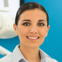
Dra. Vazquez earned her Dental Degree in the prestigious Guadalajara Autonoma University College of Dentistry, she achieved excellence in general dentistry and completed a one-year of general residency practice in the same institution (UAG 2001-2006) Dr. Vazquez then continued onto a two-year specialty-training program, where she obtained a Certificate of Advanced Graduate Study, a specialty degree in Endodontics (2005-2007) in the UAG (University Autonoma de Guadalajara) and she also attended the University of Loma Linda (L.A USA) to study for her final exam, she is a member of the Mexican Dental Association and member of the Quintana Roo Study Club. Dr. Vazquez continues with her educational courses each year.
Dra. Vazquez has been working since 2008 in one of the most prestigious dental clinics in Cancun ¨Dental Evolution Clinic¨. She has been a private practitioner in the Endodontics treatment in Cancun since 2007. She is a member of the Mexican Dental Association, the Quintana Roo Study Club and participates every year in World Endodontics Congress.
Dra. Vazquez speaks English, specializes in Root Canal Treatments and is a great handler in dental emergencies, infections, abscess and apexification.
Benemerita Universidad Autonoma de Puebla
CED. PROF. 5805980
Odontologist / Implantalogist
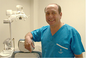
Dr. David Enriquez is a graduate from one of the most prestigious Universities in Mexico, Benemerita Universidad Autonoma de Puebla (BUAP), where he acquired his knowledge and practices as an Odontologist. Dr. Enriquez has lived in Cancun since 2005; he has been able to place himself as one of the best dentist in the city providing high-level dental treatments with full mouth rehabilitation. In 2009, Dr. Enriquez gathered and formed a team of specialized doctors and together they started the Dental Evolution Clinic project. Where he was placed as the Director of the Clinic and has maintained the tittle due to his outstanding accomplishments.
Dr. Enriquez has graduated with an Implantology Diploma through the Universidad Anahuac de Merida in June 2013, however in 2012, he was certified in a course of one year in Cancun. Since then Dr. Enriquez is able to complete both dental implant phases, the placement of the dental implant (surgically insert implant) and the rehabilitation phase (placement of abutment and crown). If dentures are needed, then the placement of titanium bars or dental structures supported by implants.
He has a wide range of implantology experience, from the placement of one dental implant, to dentures over implants and even more complex treatments such as the All-On-Four technique. He obtained through the Nobel Biocare Company an intensive introductory course on how to use the NOBEL CLINICIAN SOFTWARE after its purchase, thus making his treatments more efficient and precise.
Dr. Enriquez is an actual member of the AAID (Asociación Americana de Implantes Dentales) he keeps up-to-date by attending international congress meetings, with courses and training that prolong his knowledge on a daily base. This also keeps him informed in all dental advances available.
Without a question the assessment with his patients is outstanding. Bringing to each and everyone one of his patients his complete knowledge, dedication, professionalism, with a touch of humbleness and kindness that only Dr. Enriquez can provide.
Universidad Autonoma de Mexico (UNAM)
CED. PROF. 3445392
Orthodontic
General Dentist
In 1994 Dr. Estrada graduated from the Universidad Autonoma de Mexico (UNAM) as an Odontologist and specialized in Orthodontics at UNAM in 1999. Dr. Estrada has dedicated the last 15 years in changing smiles, not just in aesthetics but also in function, she has treated patients and concluded treatments successfully with TMJ (Temporomandibular joint and muscle disorders), as well as, patients with grinding issues. Every year Dr. Estrada attends an international congress of Orthodontics, she forms part of the AAO (American Association of Orthodontics), the World Federation of Orthodontics, Asociacion Mexicana de Ortodoncia and is certified by the Invisalign Brand 2015. Without a doubt she is one of the best Orthodontics in the Cancun area. Dr. Estrada has achieved excellent results in all or her treatments with a complete patient satisfaction.
Dr. Estrada has been in the Dental Evolution team since 2009. She has assisted and helped in a joint effort, along with other Doctors, to resolve complex dental treatments. Dr. Estrada can maintain an English conversation without any difficulty.
Universidad Veracruzana (UV)
CED.PROF. 7216590
General Dentist
Dr. De La Rosa graduated as an Odontologist from University of Veracruz in 2010. Ever since, he has dedicated his career with great devotion in General Dentistry, specializing in cosmetic treatments and mouth rehabilitation. In 2012 he became a great addition to one of the prestigious dental clinics in the Cancun area-Dental Evolution. And even now, Dr. De La Rosa has been providing great satisfactory treatments to all of his patients.
Every year Dr. De La Rosa travels to UNAM (Universidad Autonoma De Mexico) for training and to get, up-to-date, on the latest dental treatments available. You can notice his quality of work and dental abilities through all of his treatments. Dr. De La Rosa is English spoken; he is distinguished by his professionalism and the personalized treatment he brings to each and every one of his patients, making this one of his most valuable assets.
Graduate from Unam Universidad Nacional Autonoma de Mexico. (1985)
Specialty in Centro de Estudios de Postgrado en la Universidad Valle de Anahuac. (1987-1989)
Certificated by Centro de Estudios Avanzados en Odontologia.
Member Founder of the Colegio Nacional de Cirujanos Dentistas Section Riviera Maya.
Has participated in several courses on National & International levels.
Certified By Sybron, Dental Specialties Ormco, In Damon System, Passive Self-Ligating Orthodontic. (2006)
Diplomado en Ortopedia Dentofacial Otorgado por la Association Odontologica Mexicana para la Enseñanza y la Investigación. (2010-2013)
Vicepresident of Colegio Nacional de Cirujanos Dentistas Section Riviera Maya. (2012-2014).
Graduate from Unam Universidad Nacional Autonoma de Mexico 1991
Specialty in Endodontics, Universidad Tecnológica de México 1994-95
Certified by the Consejo Mexicano de Endodoncia No. 260, Marzo – 2001, first re-certification Mar 2006, second re-certification Mar 2011
President of the Colegio Nacional de Cirujanos Dentistas Section Riviera Maya Bienio 2010-2012.
Visiting Professor in Endodontics in the Facultad de Odontología de la Universidad Autónoma de Coahuila and from the Unitec.
Certified in Endodontics, Universidad Nacional Autonoma de Mexico, Fes Iztacala clinica Acatlan 1998 to 2007
Profesor Titular de Pregrado y Posgrado in Endodontics in the Universidad Tecnológica De Mexico De 1997 A 2003
Member of the Asociación Mexicana de Endodoncia
Member of the American Association of Endodontists
Member/Founder of the Colegio Nacional de Cirujanos Dentistas seccion Riviera Maya
Honoric mention in the Premio Nacional de Investigación otorgado por la AME 1998
Has published in the Journal of Endodontics
Participatión in diverse Nacional and internacional Courses.
National Conference Speaker
Graduate from Unam Universidad Popular Autonoma de Puebla UPAEP, Ced. Prof. 3788562
Congress participant 2011, 2012, 2013, presented by the Colegio de Cirujanos Dentistas de la Riviera Maya.
Member of the Colegio de Cirujanos Dentistas de la Riviera Maya
General Practice
Expert in Aesthetic Veneers
Expert in Dental Whitening.
Graduate from Unam Universidad Popular Autonoma de Puebla UPAEP, Ced. Prof. 5888261
Congress participant 2011, 2012, 2013, presented by the Colegio de Cirujanos Dentistas de la Riviera Maya.
Member of the Colegio de Cirujanos Dentistas de la Riviera Maya
General Practice
Unam Universidad Nacional Autonoma de Mexico Ced. Prof. 3705883
Congress participant 2011, 2012, 2013, presented by the Colegio de Cirujanos Dentistas de la Riviera Maya.
Member of the Colegio de Cirujanos Dentistas de la Riviera Maya
Aestetic Odontology
Clinical Assistant Professor, Department of Prosthodontics, LSU School of Dentistry, New Orleans, LA July 1, 2011-2014
Coordinator, Implant and Esthetic Fellowship Program, Department of Prosthodontics, LSU School of Dentistry July 1, 2011-2014
Dr Berron DDS has extensive knowledge and abilities in Prosthodontics & Implant Dentistry. Patients traveling from United States and Canada are able to access affordable, quality and long lasting dental implant solutions.
From a Single Dental Implant procedures to complete rehabilitation of endotolous patients requiring four or more dental implants to support fixed arches.
Instructed in the Following Didactic Courses:
Cosmetic Dentistry
Dental School Universidad Intercontinental (1981-1985, Mexico City)
Post Graduate Universidad Anahuac del Caribe. Diplomado Nutricion (2000-2001)
Practice focused in prevention
Prosthetic / cosmetic and operative dentistry
Experience:
Certified nutritionist
Dr. Alfredo Locht Office (1984-1988) Circuito Fuentes Del Pedregal En Mexico
Odontologia Integral Y Preventiva S.C. (1992-2000)
Dentaris Cancun Since 2000
Orthodontic and Pediatric specialist
One of Cancun's longest serving and respected orthodontic and pediatric dental specialists.
Graduated from UNAM (National University of México)
Post Graduated in Pediatric Dentistry. UNAM
Post Graduated in Orhodontics. UNAM
Certification: National Board Certified of Orthodontics.
Member of Mexican Dental Association.
Implant dentistry, Prosthodontics
Private practice in mexico city (1994 since 2006).
Dentaris Odontología Integral: since march 2007 until today.
Practice focused in prevention, prosthetic, operative dentistry, implant surgery and implant rehabilitation.
Education:
Bachelor of dental surgery from the Universidad Intercontinental, Mexico City (1983-1987)
Post graduate degree in integral odontology from the Universidad Tecnologica de Mexico (1992-1994)
B.t.i. Implant dentistry diploma (2001)
Endodontics with endosequence rotary instrumentation diploma (2005).
Teaching experience professor of dental anatomy and integral odontology at the Universidad Intercontinental, Mexico City (1993 – 2007)
Courses and conferences:
International ceramic symposium San Diego, California, june 2011-06-21.
Prosthodontics
School: Prosthetic Dentistry UNAM
Cirujano Dentista,
Facultad de Odontología de la Universidad Autónoma de México
Postgraduate: Prótesis Bucal Fija, Unidad de Postgrado de la Facultad de Odontología de la Universidad Nacional Autónoma de México